Maxillo-facial surgery planning and simulation
Introduction
Cleft lip and palate are among the most frequent inborn malformations.
A resulting maxillary hypoplasia can be treated by
distraction osteogenesis:
During an operation the appropriate bony part of the midface is separated
from the rest of the skull (osteotomy) and slowly moved into the ideal
position by way of a distraction device (cf. Figure 1).
Thus even large displacements over 20 mm can be treated effectively.
 |
 |
 |
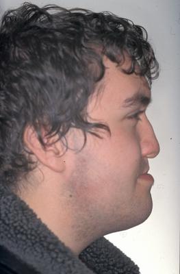
| Patient before operation |
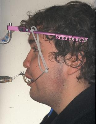
Patient with Halo device mounted |
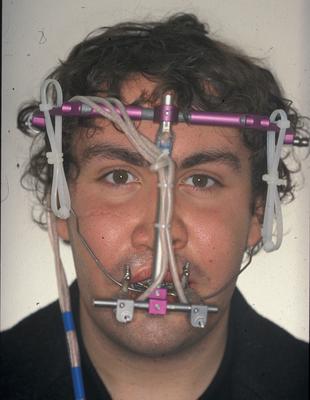
Frontal view |
| Figure 1: Maxillary hypoplasia and distraction osteogenesis
(photographies: Dr. Hierl) |
A critical point in this procedure is osteotomy design and predictions of the resulting
outcome with respect to aesthetics.
Another important question is the optimal adjustment of distraction forces,
as on one hand, one wants to pull as fast as possible, while at the other hand,
to large stresses cause unnecessary pain for the patient.
In the current clinical practice,
planning is solely based on CT scans and the surgeon's experience.
In this work package of GEMMS,
a tool chain is developed which allows the surgeon
- to specify arbitrary cuts of facial bones (pre-processing),
- to simulate the displacements of bones and soft tissue, and
- to visualize the results in an appropriate way (post-processing).
It therefore provides the possibility to predict and compare
the outcome of different surgical treatments in silico (see Figure 2).
In line with the overall goals of GEMSS, this toolchain gives
average clinicians access to
the advanced simulation technology of such surgical planning tools.
 |
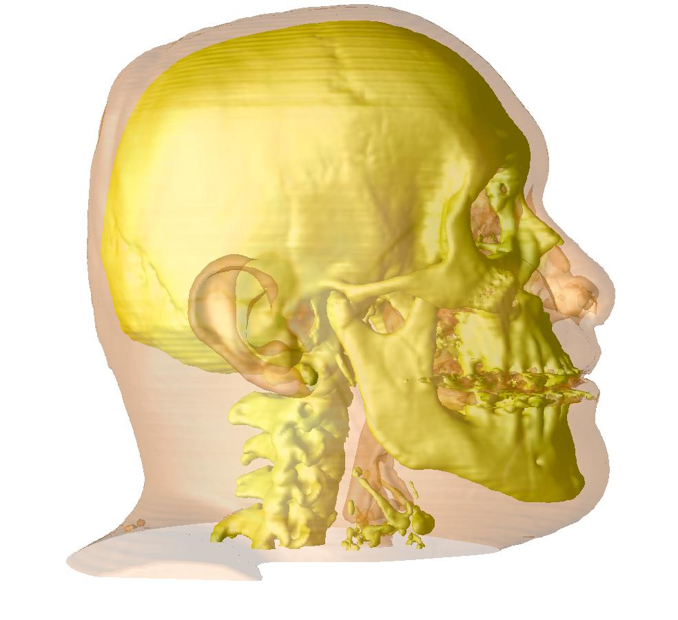 |
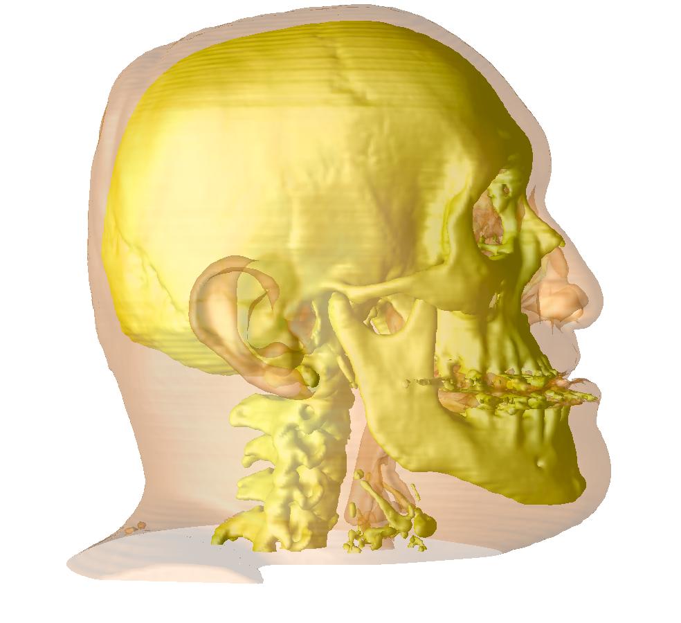
| Volume rendering of patient CT before simulation |
Volume rendering of simulated distraction |
| Figure 2. Data provided by Dr. Hierl |
The surgery planning toolchain
The chain of tasks to be solved to plan and simulate a surgery operation
includes the following steps, given a CT image of the patient:
We chose to use a loose coupling of components for solving the individual tasks,
which results in a very flexible and extensible approach.
Some of these tasks are discussed in more detail below.
We have built our virtual bone cutting tool on top of OpenDX,
which is a very flexible visualization system with a graphic programming language and sufficient
interactive power. We added some specific modules to OpenDx to realize interactive drawing of
osteotomy lines on the bone surface.
Demos are available for the cutting (3MB)
and the distraction (2.6MB) steps in the osteotomy tool.
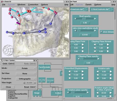 |
| Figure 3: Screenshot of bone cutting tool, based on OpenDX |
For both surface and volume mesh generation, we use the octree-based mesh generation
Vgrid and its successor
Mesh&More which is a NEC inhouse development.
Some of the algorithms are explained in [Berti2004].
We use our inhouse parallel FEM code
FEBiNA (Finite Elements for Bio-Numerics Applications)
for simulating the distraction in maxillo-facial surgery.
This code is used for linear and nonlinear FEM calculations,
where nonlinear
means as well geometric nonlinearity
as material nonlinearity (hyperelastic, viscoelastic).
Using a material model like viscoelasticity is crucial for obtaining reliable transient behavior
with respect to forces [Schmidt2004].
The final result can be visualized using volume rendering (Demo (3.5MB)).
Links and references
Related research papers
(See also the GEMSS publication list.)
- [Berti2004]
- G. Berti:
Image-based unstructured 3D mesh generation for medical applications,
in proceedings of
ECCOMAS 2004.
(download pdf).
- [Schmidt2004]
-
J. G. Schmidt, G. Berti, J. Fingberg, J. Cao, G. Wollny:
A Finite Element Based Tool Chain for the
Planning and Simulation of Maxillo-Facial Surgery,
in proceedings of
ECCOMAS 2004.
(download pdf).
Cooperation
Links to research in maxillo-facial surgery simulation
 Guntram Berti
Last modified: Tue Feb 26 14:57:41 CET 2013
Guntram Berti
Last modified: Tue Feb 26 14:57:41 CET 2013